Get in touch: (800) 727-0780


Passage 4: Hemoglobin
Hemoglobin is a vital protein found in red blood cells that is responsible for transporting oxygen from the lungs to tissues throughout the body. The structure of hemoglobin is crucial to its function and is a prime example of the relationship between protein structure and function.
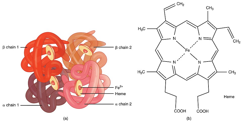
Hemoglobin is a tetramer composed of four subunits: two α-globin and two β-globin chains. Each subunit contains a heme group, which consists of an iron atom bound to a porphyrin ring. The iron atom in the heme group is responsible for reversibly binding to oxygen molecules. The globin chains fold into a globular shape, creating a hydrophobic pocket that protects the heme group and facilitates oxygen binding.
The conformational stability of hemoglobin is maintained by various intramolecular interactions. These interactions allow hemoglobin to maintain its tertiary and quaternary structure, which is essential for its function. However, certain conditions, such as changes in pH or the presence of allosteric effectors, can alter the conformational stability of hemoglobin and affect its oxygen-binding properties.
Hemoglobin exhibits cooperative binding, which is achieved through allosteric interactions between the subunits. When one subunit binds to oxygen, it induces a conformational change in the other subunits, making them more receptive to oxygen binding. This property allows hemoglobin to efficiently load oxygen in the lungs and release it in the tissues.
The oxygen-binding affinity of hemoglobin can be modulated by various factors, such as pH, carbon dioxide concentration, and the presence of 2,3-bisphosphoglycerate (2,3-BPG).
Hemoglobin can be separated and purified using various techniques, such as ion-exchange chromatography and gel filtration. These techniques rely on the differences in charge and size of the protein molecules. Ion-exchange chromatography separates proteins based on their net charge, while gel filtration separates them based on their molecular size and shape.

.svg)

